


surgical robotics development

How fast can you bring new Surgical Robotics tech to market? Know the 3Cs to rapid development.

SUMMARY
  • Speed to market has essential benefits for all top Robotic Assisted Surgery (RAS) companies – from maintaining and driving investor confidence to realising the commercial value as quickly as possible. But speed is often associated with risk.
  • Bringing new surgical robotics development tech to market quickly and confidently requires the 3Cs: Capability, Capacity and Culture.
  • A skunkworks approach provides R&D teams with all 3Cs, allowing them to focus on working without interference or interruption.
  • Our RAS leadership team will be speaking at DeviceTalks Boston on April 30, on the 3Cs and showcasing an example of a successful robotic assisted surgery device design and product development for one of our clients.

Companies that build technologies for robotic-assisted surgery (RAS) are under growing pressure to deliver new products quickly. Whether you’re a global corporation or a small startup, you need to show executives and investors that you can rapidly innovate and go to market, and you need to realize the value of investment as quickly as possible. The keys to moving fast, avoiding risks and maximizing the odds of market success? Capability, Capacity and Culture.

If you’re a longtime leader in MedTech, you undoubtedly have capabilities with a large in-house talent pool of experienced engineers, design experts and other specialists. But your development efforts can stumble if teams are stretched thin across multiple projects or are delayed by bureaucratic red tape – which are problems of capacity and culture.

If you’re a startup, on the other hand, you might have launched with a promising concept but lack the breadth and depth of capabilities, and the maturity in all three Cs. Without a broad and deep field of experts – capability and capacity – and a proven culture built on the experience of delivering many other previous projects quickly, you might struggle to meet investors’ expectations of fast results.

What’s the solution to bringing all 3Cs together? A skunkworks approach.

“A skunkworks approach enables R&D teams to focus on projects in a way that’s protected from distraction, such as internal politics,” says James Boonzaier, Deputy Head of RAS and part of Cambridge Design Partnership’s new RAS leadership team. “Too often, what we see is that key people are constantly being pulled onto other projects, or they’re spread too thin, or there are political agendas getting in the way of things. In order to get these programs running fast, you need capability, capacity and culture. Capability means, ‘Do you have the required skills across your individual engineers’ brains?’ Capacity means, ‘Are they available right now?’ And culture is, ‘Are they able to work in the right kind of context for fast, deliberate progress?’”

James-Boonzaier-e1744806299774

“In order to get these programs running fast, you need capability, capacity and culture.”

James Boonzaier | Deputy Head of RAS at Cambridge Design Partnership

A Better Approach to Rapid Surgical Robotics Development & Design

Cambridge Design Partnership’s (CDP) new RAS leadership team is headed up by Tom Brittain, James Boonzaier and Jack Hornsby. Their focus on fast delivery reflects CDP’s ongoing investment and long record of success in surgical robotics

CDP strengthens its clients’ RAS programs by enhancing capability, capacity, and culture. The company’s specialists work closely with client development teams to accelerate innovation and delivery.

“Key to working this way is being able to match highly skilled and technically capable people with the right expertise, experience, personalities, and a passion for RAS, and providing them with the right tools, conditions and working environment within which to do the best possible job,” says Tom Brittain, Head of RAS. “By collaborating closely with our clients’ teams, we’re able to operate as an extension of their own pool of people. And since we’ve been active in this sector, having worked with a number of the big players in surgical robotics, we know this approach works time and time again.”

This way of working, Tom says, enables quick development by providing organizations with turnkey access to deep technical expertise, system-level thinking and user-focused design capabilities. He notes that CDP’s success in the sector relies on a team dedicated and often exclusive to surgical robotics, with bespoke teams of experts often embedding with clients according to the unique requirements of each project.

“We’re passionate about our mission and committed to helping our clients’ RAS projects succeed,” says Tom. “And our approach works, as our track record shows. Since 2019, we’ve built four complete systems, along with multiple subsystems, for clients from the ground up. These have included both blue-sky development and remedial redesign with total system architecture development. Each of these systems has been designed and manufactured in under a year, which represents extraordinary speed in this industry. We have supported several single port trans-umbilical NPD programs, with detailed engineering of capital equipment, disposables and reposables, draping systems and more. This was made possible only by having the right capabilities on site, with the right capacity available as needed, all supported by a culture of innovation.”

Tom-Brittain-e1744807155147

“Since 2019, CDP has played a substantial role in four notable product development programs in robotic-assisted surgery, delivering system-level prototypes from the ground up. Each of these has been designed and manufactured in under a year, which represents extraordinary speed in this industry.”

Tom Brittain | Head of RAS at Cambridge Design Partnership

You need a strong foundation from which to build any RAS system

The development of any RAS system requires a strong foundation of strategy and system architecture between hardware and software, all aligned to the clinical user’s needs.

James states, “To ensure strong system architecture for our clients, we really focus on conducting structured clinical needs gathering to generate solid initial hypotheses on key requirements and constraints. Once a strong foundation for the platform architecture is created, we shift our focus to the high-risk areas.”

Risk management is vital throughout the entire process. Jack calls the approach CDP takes “targeted derisking,” adding, “we know from experience what the concept killers are and what we need to do to gain confidence that we’ve solved or avoided them. That is a key element of moving quickly. Not only that, the way in which we derisk is targeted in such a way that we’re not adding more complexity or unknowns. This enables us to iterate designs quickly, learn from each step what works and what doesn’t, and develop working prototypes at speed.”

Putting this approach into practice, CDP developed a single-port RAS prototype platform, moving from an early-stage concept to an alpha system prototype for pre-clinical trials in just seven months. Jack makes the further point that “In addition to speed, the client gets to retain all knowledge and intellectual property rights for any technologies developed for them.”

Jack-Hornsby-JH1-1

“The way in which we derisk is targeted in such a way that we’re not adding more complexity or unknowns. This enables us to iterate designs quickly, learn from each step what works and what doesn’t, and develop working prototypes at speed.”

Jack Hornsby | Deputy Head of RAS at Cambridge Design Partnership

Meet the RAS Team at DeviceTalks Boston

Building a surgical robot takes true teamwork. Success demands rapid development while meeting strict clinical and regulatory requirements. Cambridge Design Partnership continues to invest in the expertise and processes that help clients bring advanced surgical robotics to market.

The result is a comprehensive offering with unmatched expertise. Clients gain immediate access to deep knowledge in kinematics, human factors, cart design, optics, systems engineering, and disposables development. And our 26,000-square-foot Pilot Production Center quickly takes a client’s project from concept to prototype build to transfer to manufacturing.

Tom, James and Jack will be speaking at DeviceTalks Boston on April 30 about the importance of the 3Cs (“How Fast Are You? Accelerating Next-Generation Surgical Robotics”). They’ll also be showcasing a platform we developed that demonstrates our approach, going from early concept to Alpha prototype for preclinical studies in under seven months. They will be on hand at stand 735 to talk about CDP’s approach to rapid development in RAS. You can also reach out in advance and chat, or book in some time to meet them in Boston.

Connect with CDP

If you would like to discuss the content of this article, please get in touch with our RAS leadership team; Tom Brittain,  James Boonzaier and Jack Hornsby

Tom Brittain, Head of RAS
tom.brittain@cambridge-design.com

James Boonzaier, Deputy Head of RAS
james.boonzaier@cambridge-design.com

Jack Hornsby, Deputy Head of RAS
jack.hornsby@cambridge-design.com
|||

CDP advances surgical robotic technology

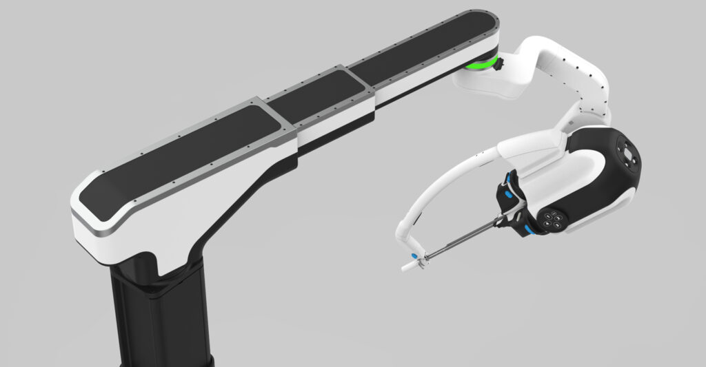
In collaboration with CDP, Titan Medical has unveiled its next-generation technology for single-access robotic-assisted surgery (RAS).

End-to-end product development partner Cambridge Design Partnership (CDP) assisted Titan Medical in designing and developing the next-generation technology for single-access RAS.

In a short time, a diverse CDP team of systems engineers, human factors specialists, industrial designers, and mechanical, electronics, and software engineers collaborated with Titan Medical to propel the development from initial concept to advanced prototype. The project is the latest example of CDP’s work in high-impact innovation in complex medical devices embodying the company’s mission to improve lives through innovation.

The Head of Robotics at CDP, says, “The starting point for this next-generation technology was the remarkable core innovations and intellectual property that power Titan Medical’s two-instrument Enos system. Titan Medical’s articulating instrument technology provides an impressive balance of dexterity and strength, allowing the surgeon to precisely maneuver them to achieve procedural tasks like grasping, suturing, and cutting. Similarly, Titan Medical’s 3D high-definition camera provides crystal clear visibility for the surgeon to be able to see the surgical site. We built on these core technologies, adding functionality for a third instrument and focusing on the usability, performance, and reliability of a RAS system.”

Cary G. Vance, President and CEO of Titan Medical, says, “CDP, with their proven record in innovation, enabled us to quickly unlock the value of our IP and convert our purpose-driven innovations and inventions into functioning prototypes. CDP has been a key partner through the years, delivering timely, high-complexity, and top-notch work. We are excited to share this achievement with them.”

By enabling surgeons to perform procedures through a single incision, Titan Medical’s technologies have the potential to reduce patient trauma and scarring and could enable patients to recover faster. This next-generation technology introduces advanced new features, such as software-enabled remote center of motion, and adds a third dexterous instrument to allow greater procedural flexibility and enhanced surgeon control. Innovations in the design allow a more compact footprint and lighter weight than current systems, taking up less space, making it easier to maneuver in space-constrained operating rooms, and providing open access to the patient.

The Head of US Office at CDP, adds, “Ultimately, the focus needs to be on the patient and on enabling the surgical team to achieve the most successful surgical outcomes safely and effectively. A key insight that fed into this development was recognizing that many RAS systems crowd the bedside and impede access to the patient. Sometimes you can barely see the patient under the robotic arms! We saw an opportunity to create a patient cart with a minimalistic and open architecture that is easy to work with and work around. Our work in advanced medical devices, particularly RAS technologies, continues to be one of the highlights of CDP’s 27-year history. This milestone is a proud moment for us, and we believe this technology will be a benchmark for single-access surgery.”

Concluding that, “Designing highly capable RAS systems means carefully navigating challenges and compromises such as performance and safety. Our approach resulted in an advanced and simple-to-use system that finely balances those difficult compromises. We delved deep into Titan Medical’s extensive intellectual property portfolio to be able to develop the best of all available technologies and bring our client’s valuable know-how to life. When working with CDP, our clients get access to our proven history of innovation, our nimble processes, and highly capable teams. This project is a great highlight of our capabilities embodied in one deliverable.”

“CDP has been a key partner through the years, delivering timely, high-complexity, and top-notch work. We are excited to share this achievement with them.”

Cary G. Vance, President and CEO of Titan Medical

WHITEPAPER

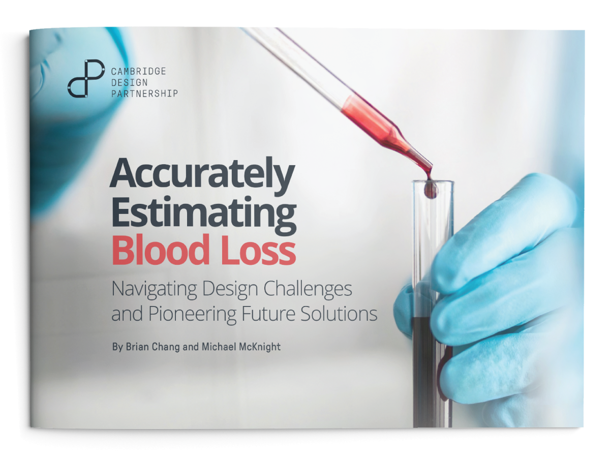
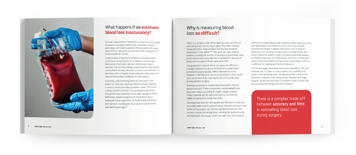
Accurately Estimating Blood Loss

Navigating Design Challenges and Pioneering Future Solutions

BY BRIAN CHANG and MICHAEL MCKNIGHT
||CDP whitepaper - Accurately Estimating Blood Loss
web_body_Accurately-Estimating-Blood-Loss

Significant advancements in surgery, such as the emergence of surgical robotics and augmented reality (AR)-assisted instrument navigation, have revolutionized patient care. Despite these innovations, there are still critical needs that technology has yet to fully address. One such need is the accurate and easy estimation of blood loss during surgery. The lack of a comprehensive solution for real-time monitoring of blood loss poses serious risks to patient outcomes.

In this article, we will explore the challenges of estimating blood loss (EBL), evaluate the advantages and disadvantages of various novel solutions, and discuss the key factors necessary for achieving more accurate, timely, and insightful monitoring.

Contact Us

At CDP, we integrate seamlessly with your team to accelerate your project’s development.
To discuss how we can help you, get in touch.

|||||||||||||

Single use endoscopy: A greener solution?

This article illustrates why developers of single-use endoscopes should consider sustainability concerns. It also presents counter-intuitive potential benefits for this sector and outlines Cambridge Design Partnership’s recommended framework for designing a greener solution.

Bronchoscopy, duodenoscopy and arthroscopy are just a few of the procedures being served by a growing device market segment: single-use endoscopes. Development in this space is active, with companies such as IQ Endoscopes and Pristine Surgical seeking to join market leaders like Boston Scientific, Olympus, and Ambu. With the size of its addressable markets and technical opportunities, I can see why this medical visualization segment is expected to grow significantly, with projected sales reaching $710 million in 2024 and potentially soaring to $3 billion by 2033.1

Clinical and Care Provider Benefits

There are multiple clinical and stakeholder benefits of single use endoscopy, with the primary one being reducing infection risks. For example, reusable bronchoscopes are particularly hard to disinfect due to their long, narrow channels, which have historically led to notable rates of infection and hospital readmission2. For duodenoscopy, infection from ineffective decontamination of reusable scopes is so concerning for the FDA that they issued a communication to support the transition to fully or partially single-use versions.3

Removing the need for disinfection can also deliver benefits to healthcare providers. It will simplify device handling workflows by reducing user steps, eliminating maintenance, and requiring fewer personnel, capital equipment, facility space, quality processes, and training typically needed for disinfection.

web_body_endoscope-disinfection

As a result of these eliminations, the transition to single-use endoscopes is also expected to reduce total costs for certain procedures, as highlighted by some manufacturers. This shift could also improve provision for smaller population centres and future care pathways, such as the increased adoption of diagnostic clinics and ambulatory surgical centres.

Addressing Sustainability Concerns

While the benefits of single-use scopes appear clear, there are concerns about sustainability. The increased material usage and disposal of electronic components after a single use may seem to contradict sustainability goals, such as UK’s National Health Service 2045 net zero target for indirect emissions.

Can we reconcile the use of single-use disposable endoscopes with sustainability?

This question can be tackled from two angles:

The Strategy Angle

Given the contamination risks associated with reusable endoscopes, it’s understandable to question the emphasis on sustainability. Would you subject a loved one to a procedure with high risks of hospitalization and death, just to be environmentally friendly?4 Considering this, some might think that sustainability can be sidelined.

However, this approach doesn’t address the concerns of medical equipment buyers and other stakeholders – with evolving environmental policies and competition from the “greenest” manufacturers, ignoring sustainability could be costly. Moreover, our experience shows that designing for sustainability can bring commercial advantages, such as lower manufacturing costs and more streamlined supply chains.

Therefore, innovators of single-use endoscope should not become complacent about sustainability. Embracing design for sustainability is crucial to stay ahead of future policy and competitive forces.

The Measured Greener Angle

Endoscope innovators looking to incorporate sustainability thinking for their solution may wonder if single-use endoscopes can ever be greener than reusable versions. Surprisingly, the answer is “yes”, in some cases, single-use endoscopes might be better for the environment.

This seems to contradict the traditional “Reduce, Reuse, Recycle” principle. However, you cannot solely rely on the 3 R’s and have a narrow perspective. To accurately measure environmental impact, a comprehensive and systematic approach is necessary. Life Cycle Assessment (LCA) is a valuable methodology for this purpose.

Life Cycle Assessment

Life Cycle Assessment (LCA) involves assessing the environmental impacts associated with all stages of a product’s life cycle, including material usage, energy consumption, transportation and other metrics from production to end of use. By using specialized databases, LCA estimates environmental factors such as carbon footprint, water usage, resource use, and toxicity levels.

CDP-flow_product-footprint
Lifecycle steps analysed for assessment

With this method, it is possible to conduct an impact analysis of a new product in comparison to an existing one to determine if the new proposition is more sustainable.

While we understand the limitations of LCA calculations, such as the accuracy of inventory data, challenges in addressing unknowns, complex transportation and establishing boundaries, it remains an appropriately credible approach with guidance available in the ISO 14044 standard.

Consider the comparison between single-use endoscopes and reusable ones in terms of sustainability factors, as outlined in Table 1.

Positive Sustainability Factors of Single-Use Scopes Negative Sustainability Factors of Single-Use Scopes
Reduced material due to lack of need to withstand multiple uses and decontamination steps Increased production of scopes and packaging
Decreased energy, water, and material usage as well as reduced need for personal protection equipment, due to elimination of decontamination cycle for each scope use Higher transportation requirements for the increased number of scopes
Elimination of impacts for maintenance effort Increased scope disposal
Reduced hospital re-admission due to reduced infection risks

The information in the table above might suggest that a reusable scope could be greener. However, conducting an LCA would be needed and can often yield surprising results.

Case Studies

An analysis published in the American Journal of Environmental Protection illustrated that the material and usage impact of decontamination of reusable scopes is so significant that Ambu’s single-use scope, the aScope 4 Cysto, has a lower carbon footprint when LCA-calculated.5

In another analysis where the effects of hospital re-admission were considered, Boston Scientific claims a 65% greenhouse gas reduction with their SpyGlass™ DS Cholangioscope, which incorporates a single-use disposable scope as part of the solution.6

While these examples show the potential for greener practices, it’s important to acknowledge that achieving sustainability may be challenging in other scenarios. Nevertheless, the key point is about environmental impact reduction, which is crucial for safeguarding the future market and conveying a compelling message to stakeholders.

web_body_Ambu-aScope-4-Cysto
Image: Ambu® aScope™ 4 Cysto. https://www.ambu.co.uk/endoscopy/urology/cystoscopes/product/ambu-ascope-4-cysto

How to Make a Single-Use Scope Greener

For the successful sustainability-driven evolution of an existing single-use endoscope or the development of a new one, it is essential to have a structured process that provides essential market-related insights to the development team. This process should encourage creative exploration and enable credible evaluation of sustainability decisions, while ensuring clinical and commercial objectives are met.

Cambridge Design Partnership’s Approach

With our extensive experience in developing more sustainable products, including medical devices, the following outlines our approach, specifically related to this segment.

Prepare

Research & Define: Understand the environmental impacts that are important to your customers, such as carbon footprint, total waste, toxicity, or a combination of these factors.

Establish: Identify which endoscope and use cases you would like to be greener than.

Target: Define the extent to which you aim to improve environmental performance for each metric.

Equip: Gain an understanding of all design techniques for sustainability, going behind the conventional “reduce, reuse, recycle” approach.

Create

Imagine: Explore various design architectures for partially or fully disposable scopes and evaluate the inclusion of non-essential features in the design.

Explore: Identify potential waste management partners to find single-use scope recycling solutions.

Calculate: Conduct rough LCAs during the design phase using user-friendly tools to guide decision making.

Deliver

Test: Gather user feedback in real-world settings to validate the proposed design.

Check: Ensure that the design meets or surpasses performance, usability, cost, and stakeholder requirements to create the right product.

Sharpen: Once closer to finalizing a design, perform a more detailed LCA using ISO 14044 guidance and iterate as needed to achieve targets.

Conclusion

Single-use endoscopes is an exciting and growing domain with opportunities for developing new products to target new indications, offer cost reductions for care providers, improve hospital workflows and, critically, achieve better clinical outcomes by mitigating infection risks.

While pursuing these goals, innovators should not overlook the potential to create more sustainable solutions, which can yield environmental benefits, deliver commercial opportunities, and ensure market protection.

Addressing sustainability requires a departure from the conventional “reduce, reuse, recycle” mindset and a shift toward an informed, creative, and data-driven approach to developing sustainable offerings.

Connect with CDP

At Cambridge Design Partnership, our proven track record in enhancing the sustainability of medical devices can help you meet both regulatory and market demands.

If you have any questions about the content of this article, please contact us.

|||

The importance of education and innovation in WOC nursing – Reflections on WOCNext 2023

For 10 years, I’ve been a critical care nurse in the ER and ICU. My colleague, Nicki Sutton, Head of Insights and Strategy and I recently attended the WOCNext 2023 conference in Las Vegas, Nevada – the first I’d been to.

Perhaps a wound, ostomy, and continence (WOC) conference would seem an odd choice for a critical care nurse, devoid of ventilators and vasopressin, and in that light, I guess it is. However, while I am still a practicing ICU nurse, I also work for Cambridge Design Partnership, an end-to-end innovation partner with expertise in product development from idea to production.

So, getting to learn more about the nuanced world of WOC certainly helps inform the clinical perspective I can bring to our clients as we work to help them develop new and novel products to heal patients in a variety of ways.

web_inline_Erik-at-WOCNext2023

Our Insights Research Associate, Erik Andersen, at WOCNext2023

As a non-WOC nurse, I must say: I am hugely impressed by the skill and the art that is WOC nursing. Especially, the art of being able to perform astute assessments on skin tones that vary so greatly and being able to ‘read’ the stoma and peristomal skin to overcome the convexity challenges, much like a golfer ‘reads the green’ to sink the putt.

I’ve always respected the knowledge that my WOCN colleagues bring to the table, but frankly, it’s easier to apportion patient care responsibilities in a hospital setting. Even though I try to help with wound care and appliance changes when I can, I don’t always get to appreciate the total value-add they bring to their patients. At WOCNext, it was a different story: the sheer breadth and depth of knowledge that the presenters and attendees possessed was astounding.

Aside from the fact that I still have a lot to learn about wound care, stoma management, and incontinence interventions, there were a number of interesting themes that we were able to pick up on throughout the presentations and discussions:

We’re losing the WOC numbers game

While it’s true of nursing across the board, many folks highlighted the systemic lack of qualified or certified WOC nurses nationally which makes satisfying the needs of their patient populations exceedingly difficult. This scarcity seems to be much more evident in home care settings where resources are already sparse, and now making both access to patient-appropriate products and WOC nurses a huge struggle.

We need to extend education to non-WOC nurses 

The second thing that struck me was how much WOC-related knowledge simply doesn’t make it to the bedside nurses, with many people pointing to a lack of education and awareness about wound care best practices and appropriate products held by non-WOC nurses. It seems as though there is an incredible degree of knowledge about these topics that don’t quite reach the broader nursing population, myself very much included.

There’s an opportunity for innovation

As a healthcare innovator, this shows me there is potential opportunity for manufacturers developing healthcare products, specifically those in the wound care and ostomy space, to understand the stark knowledge gap between WOC and non-WOC nurses, and design systems and products that can help upskill non-WOC nurses to offer better coverage of WOC management.

By no means am I suggesting a WOC nurse could be replaced, but rather to the contrary: they’re at a numeric disadvantage and already working hard to cover the patients that need them.


So how can we democratize WOC care so that the lack of available specialists doesn’t have such a negative impact on the patients requiring their care? For instance, maybe there’s room to lean on a technology aid to remove some of the subjectivity that an artful WOC nurse can account for, but that the less specialized nurses among us, like me, can leverage to do a better job with assessments or appliance management.

That’s the kind of work we can support at CDP, where we have been innovating in the healthcare industry for over 25 years. We have experts across the product development pathway: from patient and clinician research, to design and engineering, to usability testing and validation, all blanketed under our ISO 9001 and 13485 certifications for medical device development and manufacture.

We aim to improve lives through innovation much like our clinical colleagues, aim to improve lives through healthcare delivery. One such example is the work we’ve done with ostomy product innovation start-up, Ostique. Over four months, we supplied the technical rigor to help them develop a concept for a unique ostomy appliance and build prototypes of the device for user feedback. Today, having successfully brought their product to market, Ostique’s revolutionary pouch covers are available in a range of colors and skin tones to meet patients’ needs.

The need for improved healthcare and delivery is there – we could see it everywhere at WOCNext 2023 – and we’re here to help you address it. To discuss how we can help your organization take your product to market, chat to the team today

Neurodegenerative conditions|||||

Neurodegenerative conditions: turning a corner to better treatment?

Pace is accelerating for tackling neurodegenerative diseases. Can we unlock better treatment? Can we reach a cure?

Ageing populations face neurodegenerative conditions, such as Alzheimer’s Disease, Parkinson’s Disease, Motor Neurone Disease, Multiple Sclerosis, and others. These impact an estimated 60 million people worldwide, equivalent to the current UK population.

Whilst each condition has different mechanisms of neurodegeneration, they all have something in common: prognosis is bleak, treatment is limited, and there is no cure.

However, after decades of research, there has been a series of breakthroughs. Here, we focus on two areas of progress: how treatments have moved on and hope for the future.

The rise of RNA-based therapeutics 

The effective development of RNA-based vaccines during the COVID-19 outbreak catapulted RNA-based therapeutics into the spotlight. Whilst theoretical knowledge of RNA therapy has existed for over 30 years, the bulk of associated FDA approval for treatments involving the nervous system has occurred in the last decade(1).

A major advantage of RNA-based therapy over conventional small molecule and protein-based approaches is its high specificity and precision, resulting in a more targeted approach to treating disease with specific gene mutations or overexpression.

However, to devise effective RNA-based therapeutics, the genetic hallmarks of the neurodegenerative disease of interest must be known.

Motor Neurone Disease (MND) is one such condition where specific mutations in the SOD1 gene have been identified and in this case, in two per cent of diagnosed cases.

A recent breakthrough in phase three clinical trials targeted this gene using the drug Tofersen. Tofersen, developed by Biogen, directly interferes with the faulty overproduction of SOD1. After six months, patients had a reduction in SOD1 levels, and after 12 months the same patients reported better mobility and lung function(2,3). Although patients with SOD1 mutations only represent two per cent of those living with MND, these trials provide ‘proof of concept’ that similar gene therapy-based approaches may help other forms of the disease.

Another pioneering strategy, developed by Atalanta Therapeutics and Genentech, focuses on a technology called branched siRNA (small interference RNA). This is a type of molecule that helps regulate gene expression by binding to a complementary messenger RNA, which in turn can encode the gene of interest.

Branched siRNA uses novel RNA interference nucleotide technology to suppress the activity of genes that function abnormally, such as mutations. This slows the progression of the disease or stops it altogether.

It is hoped this approach can be applied across multiple neurodegenerative diseases, including Parkinson’s Disease, Huntington’s Disease and Alzheimer’s Disease.

Although testing is still in the pre-clinical stage, the branched siRNA platform aims to enable RNA interference to be deployed as a therapeutic approach throughout the brain and spinal cord. This overcomes the long-standing challenge of achieving adequate distribution within the central nervous system (CNS) to ensure the therapeutic agent reaches the nervous tissue(4,5).

Progress in non-RNA therapeutics 

Non-RNA therapeutics for neurodegenerative conditions also continue to progress. Examples include the monoclonal antibody Donanemab, developed by Eli Lilly. Phase three clinical trials showed it to slow clinical decline by 35% in patients with Alzheimer’s Disease, compared to a placebo(6).

Effective delivery remains a major challenge  

One of the main challenges in developing RNA therapeutics, and therapeutics for the brain in general, remains the efficiency of its delivery to the target tissue.

To treat neurodegenerative conditions, the therapeutic agent aims to reach the CNS. The presence of the blood-brain barrier (BBB), a cell-formed wall separating the bloodstream and the CNS, makes it difficult to deliver drugs. The BBB’s almost impermeable characteristics allow very few molecules to cross and make systemic drug delivery less efficacious.

There are two common approaches to overcome this: re-engineering the therapeutic agent to make it compatible with BBB permeability or bypassing the BBB altogether.

Re-engineering the therapeutic agent

This typically involves chemical modification of the drug (e.g., from water-soluble to lipid-soluble molecules) to enable passive diffusion through the BBB. Another approach is to design drug carriers that mimic the structure of endogenous molecules (e.g., monosaccharides, hormones) to activate carrier-mediated transport or nanocarriers(7,8). Both approaches add complexity to manufacturing.

Another cross-BBB approach is Focused Ultrasound (FUS), where high-intensity sound waves temporarily disrupt the BBB to enable drug-loaded microbubbles to enter the CNS9.

Bypassing the blood-brain barrier 

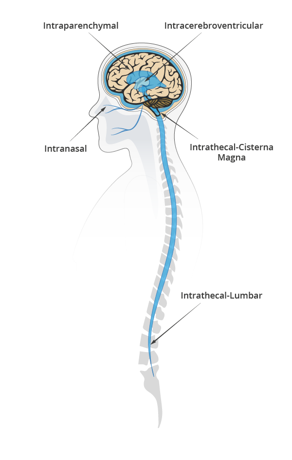
Bypassing the BBB can save time and effort in formulation by using a range of therapeutic agents not constricted by size or BBB compatibility. Of its three most common types of delivery: intraparenchymal, intranasal, and cerebrospinal fluid (CSF) delivery; the latter is often the favored approach, due to lower clinical complexity10.


Evaluating CSF delivery routes 

CSF delivery most commonly include intrathecal (IT) or intraventricular (ICV) routes.

IT involves an injection either on the lumbar or a cisterna magna region to deliver the drug and let CSF pulsatile flow support the distribution of the therapeutic agent in the brain and spinal cord.

ICV is more invasive. It involves two surgical interventions, one to place a catheter connecting the cerebral ventricles to the injection port at the top of the skull and one to remove the catheter.

To date, ICV has two approved drugs (Rituxan for CNS Lymphoma, and Brineura for Neuronal Ceroid Lipofuscinoses type two). IT lumbar injection has one (Spiranza for Spinal Muscular Atrophy) and plenty more in clinical and pre-clinical stages across a spectrum of neurodegenerative and neurological diseases(11). Irrespective of the approach, the trend is clear: less invasive, lower dosage, and targeted delivery is the way to go.

In the race to show safety and efficacy with either invasive or non-invasive approaches, all solutions will have to be patient-centered.

A new dawn for the treatment of neurodegenerative diseases  

The complexities of neurodegeneration have long frustrated scientists and clinicians alike, despite decades dedicated to studying its diseases, aetiologies, and treatments. However, we are making more rapid and more significant progress.

We have some way to go, but we mustn’t overlook the magnitude of these milestones. New therapeutics and delivery techniques are paving the way to more effective and efficient treatment.

By increasing our understanding of genetic hallmarks of the diseases, and using tools such as AI in drug discovery, we can unlock faster pathways to RNA-based treatments. Similarly, by finding innovative ways of demonstrating the safety and efficacy of delivery methods, such as modeling, we can edge closer to less invasive procedures and lower dosages to minimize potential side effects.

We need more research, more awareness, earlier diagnosis, and a better understanding of risk factors to enable prevention and earlier intervention.

But we are now getting closer to better treatment and one day finding a cure.


References 
  1. http://nectar.northampton.ac.uk/16015/1/Anthony_Karen_RNAB_2022_RNA_based_therapeutics_for_neurological_diseases.pdf
  2. https://www.sheffield.ac.uk/neuroscience-institute/news/promising-mnd-drug-helps-slow-disease-progression-and-benefits-patients-physically
  3. https://www.nejm.org/doi/full/10.1056/NEJMoa2204705
  4. https://www.gene.com/stories/pioneering-novel-therapeutics-in-neuroscience
  5. https://www.nature.com/articles/s41587-019-0205-0
  6. https://clinicaltrials.gov/ct2/show/NCT04437511?term=TRAILBLAZER-ALZ&cond=Alzheimer+Disease&draw=2&rank=3
  7. https://www.ncbi.nlm.nih.gov/pmc/articles/PMC8905930/
  8. https://ijponline.biomedcentral.com/articles/10.1186/s13052-018-0563-0#:~:text=Modification%20of%20the%20drug%20to,capable%20of%20crossing%20the%20BBB.
  9. https://clinicaltrials.gov/ct2/show/NCT03321487
  10. https://www.frontiersin.org/articles/10.3389/fnagi.2019.00373/full
  11. https://www.ncbi.nlm.nih.gov/pmc/articles/PMC9305158/


Connect with CDP

For more on how to advance RNA therapeutics and targeted CNS drug delivery for neurodegenerative diseases, contact Cambridge Design Partnership.

Our brains in space|||||

Our brains in space: The next giant leap

Fifty-two years after the first lunar landing, NASA’s program Artemis prepares to land on the moon.It’s a major step towards future space explorations and Mars missions – one that’ll see women in deep space for the first time.

The technology being developed includes the next generation of rockets, space suits, lunar landers, and gateways for travel into deep space. However, we’re yet to solve one of the most fundamental aspects of long-duration space travel: how to keep our brains alive.

The human brain is a remarkable feat of physiological engineering. It has sensing systems, self-regulating mechanisms, valves, dampening features, adaptable form, and even a set of complex networks that developers of the most advanced AI can only dream of. Yet, we still know surprisingly little about the interactions and boundaries of these systems and how they respond to space travel.

In this article, I explore how mathematical modeling is improving our understanding of the brain, successfully predicting the existence of anatomical features no one’s identified before – with learnings for Mars missions and the diagnosis and treatment of conditions such as Alzheimer’s disease.

What happens to our brains in space?

The human body is adapted to operate with Earth’s gravity. In space, microgravity (reduced gravity) causes fluids in the body to shift. This is most noticeable in the brain, where microgravity increases fluid volume, putting pressure on the nervous tissue.

Having a rigid cranium is good news for many reasons, but holding excess fluid isn’t one of them. Luckily, the increased intracranial pressure isn’t high enough to be immediately fatal. Still, it can introduce chronic knock-on effects with long-term implications.

Immediate brain-related effects experienced by astronauts due to increased intracranial pressure and fluid movement in the inner ear include headaches, dizziness, nausea, temporary blurred vision, and disturbed perception and spatial awareness. During space flight, the brain’s vestibular (orientation) system adapts to the new environment, reducing some of the effects. But others persist or appear in different forms, such as sleep disturbances.

After returning to Earth, astronauts who’ve taken long-duration flights have had one or more of these effects:

  • visual impairment – ophthalmic changes due to excess pressure in the optic nerve
  • brain ‘swelling’ – changes in ventricles (fluid-filled cavities) and grey and white matter (brain tissue composed of nerve fibers and nerve cells)
  • pituitary gland shrinkage – downsizing of the hormone-secreting gland that helps regulate body functions, including blood pressure and kidney function.

There’s a correlation between longer-duration space flight and increased severity of post-flight effects. These effects can be present long after the mission ends, and some can cause irreversible damage, for example, visual impairment.

Why does this happen?

The brain is protected for changes in gravity by a surrounding fluid called cerebrospinal fluid (CSF), a non-permeable layer called dura mater where CSF is contained, and one-way valves called arachnoid villi on the dura mater where CSF exits.

CSF is responsible for compensating changes in pressure, such as those created by the arteries or changes in posture. It’s also involved in cellular waste removal from the brain during sleep, ‘cushioning’ the brain to prevent trauma, and maintaining the best possible environment for the brain, for example, optimizing ion concentration levels to prevent cell death.

Dura mater is the ‘sac’ that contains the CSF. It plays a pivotal role in ensuring CSF exits the cranial and spinal spaces in a controlled way through the arachnoid villi.


web_mathematical-modelling-our-brains-in-space-diagram-1

As gravity lessens, important events take place which exert pressure in the brain:

  • CSF in the cranium is no longer free to flow towards the spinal spaces. It concentrates mainly in the cranial space, increasing intracranial pressure.
  • The spinal dura mater, usually relaxed and undisturbed, becomes compressed by other fluids and nearby organs as they shift upwards.
  • Upward movement of the brain and spinal cord stem obstructs one of the main CSF exit routes, the superior sagittal sinus (SSS, composed of arachnoid villi). This prevents relief from increasing CSF pressure in the brain.

Gravity influences low-pressure systems most, so blood flow in the veins is affected, causing ‘pooling’ where blood accumulates because of poor circulation. This matters because cerebral veins lack valves, relying on CSF to ‘push’ venous blood forwards. There’s also movement of organs in the upper body and extracellular fluid, putting extra pressure on the spinal cavity and the veins in the head and neck. Other effects play a role in what’s known as ‘fluid shift. These include osmotic pressure changes (fluid motion between areas of different solute concentration levels), variation in heart rate and respiration, and changes to lymphatic dynamics (the movement of a fluid, known as lymph, from tissues and organs).

How do we model the brain?

Scientific efforts focus on three approaches:

  • microgravity testing – space mission and Zero-G flights with, for example, lower-body negative-pressure gravity suits, which aim to ‘pull’ fluid away from the upper body
  • on-Earth simulated testing – bed-tilt tests to measure physiological changes during bed rest
  • mathematical modeling.

Given the promise of mathematical modeling in predicting difficult-to-measure behaviors, such as epidemiological events like the ongoing COVID-19 pandemic, it’s worth exploring further.

Modeling the brain and spinal cord in microgravity

As part of my research, I’m investigating the relationship between pressure fluctuations in the brain and spinal cord and their impact under different gravitational conditions.

Using mathematical modeling, I outline the relationship of a single arterial root vessel and its blood flow with the next generation of vessels. This root vessel branches into two vessels, and each of those to another two, and so on. Each generation carries information of the preceding vessel from which the rest of the parameters can be derived. The arterial branching continues until reaching the capillaries (tiny vessels where cellular nutrients and waste are exchanged), before converging again to complete the venous tree.

After the blood vessels are described, I add the CSF spaces in the cranium and spine. These spaces allow CSF to interact with the blood vessels, which I can study for behavior under different physiological and gravitational conditions

web_mathematical-modelling-our-brains-in-space-diagram-2-1

The model accounts for several key features of cerebral circulation, including autoregulation (the ability of blood vessels to expand and contract to metabolic demands and pressure changes), CSF exit routes (arachnoid villi), and gravitational components. As I add gravity to the model, I can test different postures, for example, lying flat on the back (supine).

By mathematical modeling, I successfully predicted the existence of anatomical features that had never before been identified – arachnoid villi in the spinal dura mater. These additional exit routes appear to be a crucial element to relieve CSF pressure under different gravitational conditions. The model showed that the cranial and spinal exit routes work together to keep CSF pressure fluctuations stable, ensuring undisturbed cerebral blood flow.

web_mathematical-modelling-our-brains-in-space-diagram-3

On Earth, standing allows CSF to shift downwards, lowering pressure inside the cranium, which causes the cranial exit route to close. The spinal exit route can work as the main exit route, but it also has to deal with increased pressure outside the spinal cavity, such as the interstitial fluid (fluid between cells) of surrounding organs. If the external pressure is greater, the spinal exit route closes, and the pressure inside the cranium increases until the cranial exit route opens again. A supine position allows exit through either the cranial or spinal routes.

In microgravity, the upward shift of fluid causes CSF pressure in the cranium to increase, whether standing or supine. This can be modeled using a bed-tilt test where the body is positioned at a negative angle. The spinal exit route closure intensifies CSF intracranial pressure because of increased pressure from other fluids and organs on the spinal cavity. The combined effect forces the cranial exit route to open to stabilize pressure fluctuations. However, recent research suggests the upward movement of the nervous tissue (brain and spinal cord) may block the main cranial exit route (the SSS). Blocking the SSS in the model leads to sustained intracranial pressure, resulting in irreversible consequences, which we see in space missions.

I also found that cerebral autoregulation plays a key role in overcoming gravitational changes in space and on Earth. Where autoregulation is absent or abnormal, constant blood isn’t delivered adequately to the brain, compromising oxygen to the brain. This can cause a range of conditions, including hypoxic events, where lack of oxygen leads to impaired or permanent nervous tissue damage.

All of this means that the brain, on its own, isn’t sufficiently adaptable to prevent tissue damage in microgravity environments. Sustained high intracranial pressure needs a path of least resistance to relieve the brain tissue of excess fluid. Extending space missions without fully understanding this behavior poses a significant risk.

The next stage of my modeling involves testing acute postural changes in combination with chronic physiological conditions, such as aging, to further analyze the effects I observed.

Why does this matter – and what can we do about it?

Beyond space exploration, understanding fluid movements in the brain and spinal cord is critically important to better diagnose and treat conditions such as Alzheimer’s disease, hydrocephalus (a life-changing and often fatal condition due to excess of CSF volume), spinal syringomyelia (a fluid-filled cyst disorder in the spine), stroke, and Multiple Sclerosis. Until we understand how these mechanisms can cause permanent damage, it’s vital to keep analyzing abnormal CSF-cerebrovascular behaviors under any gravitational conditions.

On whether we can go to Mars, it’s possible, but landing on Mars (roughly seven months of space travel each way) would increase the risk of developing long-term health conditions, and as the mission extends, so does the likelihood and seriousness of those risks.

However, by pursuing an understanding of those risks, we’ll understand our physiology better. What adaptations could be expected of the human brain under any gravitational condition? What can we learn from other mammals overcoming significant gravitational challenges, such as bats and giraffes? Can an understanding of human brain adaptations help us treat neurological or neurodegenerative conditions?

As we aim for ever more challenging space missions, mathematical modeling can help us characterize the boundaries of human physiology – and take the next giant leap in understanding our brains.


References
  • Kramer, L.A., Hasan, K.M., Stenger, M.B., Sargsyan, A., Laurie, S.S., Otto, C., Ploutz-Snyder, R.J., Marshall-Goebel, K., Riascos, R.F. and Macias, B.R., 2020. Intracranial effects of microgravity: a prospective longitudinal MRI study. Radiology295(3), pp.640-648.
  • Li, K., Guo, X., Jin, Z., Ouyang, X., Zeng, Y., Feng, J., Wang, Y., Yao, L. and Ma, L., 2015. Effect of simulated microgravity on human brain gray matter and white matter–evidence from MRI. PloS one10(8), p.e0135835.
  • Petersen, L.G., Lawley, J.S., Lilja‐Cyron, A., Petersen, J.C., Howden, E.J., Sarma, S., Cornwell 3rd, W.K., Zhang, R., Whitworth, L.A., Williams, M.A. and Juhler, M., 2019. Lower body negative pressure to safely reduce intracranial pressure. The Journal of physiology597(1), pp.237-248.
  • Roberts, D.R., Albrecht, M.H., Collins, H.R., Asemani, D., Chatterjee, A.R., Spampinato, M.V., Zhu, X., Chimowitz, M.I. and Antonucci, M.U., 2017. Effects of spaceflight on astronaut brain structure as indicated on MRI. New England Journal of Medicine377(18), pp.1746-1753.
  • Roberts, D.R., Asemani, D., Nietert, P.J., Eckert, M.A., Inglesby, D.C., Bloomberg, J.J., George, M.S. and Brown, T.R., 2019. Prolonged microgravity affects human brain structure and function. American Journal of Neuroradiology40(11), pp.1878-1885.
  • Sanchez-Cazares, K E 2018, ‘Mathematical Model of the Cerebral Circulation and Distribution of Cerebrospinal Fluid’, PhD thesis, Imperial College, London.
Incisive action: Cutting the carbon footprint in surgery|

Incisive action: Cutting the carbon footprint in surgery

Hear us out: the pandemic has stretched world health services to their limits, but it may also be paving the way toward a greener future for healthcare.

When thinking of healthcare today, you probably picture the huge pressures on overworked healthcare staff and the scramble for hospital beds. What you may not have thought about is that hospitals in many countries have adopted innovation that inadvertently introduced ‘greener’ treatment. For example, the need to perform ‘virtual’ consultations has reduced patient travel to and from practices. In April 2020; within weeks of COVID-19 hitting the UK, 71% of all GP visits were remote, compared to 25% in April 2019.

A single operation can have the same carbon footprint as driving 2,273 miles in an average sized gas-powered car.

Before COVID-19, the UK’s National Health Service (NHS) produced 27 million tons of CO2 equivalent annually, which accounted for 5% of all UK carbon emissions. To combat this, in October 2020 the UK government announced plans for a greener NHS: net zero carbon emissions directly from the NHS by 2040, and its supply chain by 2045.

In the context of COVID-19, this is an ambitious goal even if we were able to sustain the kind of CO2 emission drops witnessed during lockdowns. The forced shutdown of elective surgery may have reduced hospital carbon footprints, but this has been at the expense of patient care and can’t continue. Further ahead, the NHS will be caring for an increasingly ageing population, putting demands on provisions which will lead to increasing energy and resource consumption.

The operating theater has extensive electricity needs, powering equipment, heating, ventilation, and air conditioning, and is three to six times more energy-intensive than the rest of the hospital. This electricity reliance coupled with anesthetic gas and the need for single-use equipment has a significant carbon footprint. Chantelle Rizan, a Fellow of the Centre for Sustainable Healthcare and currently undertaking a PhD to identify carbon hotspots in surgery, found that a single operation can have the same carbon footprint as driving 2,273 miles in an average sized gas-powered car.

So, aside from upgrading hospital buildings and moving to renewable energy supplies, the UK government must explore ways to make surgical practice more sustainable in order to hit the NHS net zero targets. This won’t be easy.

Virtual clinics have helped with triage (deciding severity and service allocation) and surgical follow-ups, but it’s difficult to plan surgery without examining the patients face-to-face. Any changes must avoid extra red tape and be economically viable for healthcare services. Advances may have trade-offs between short-term losses (retraining) and long-term gains (reducing hospital stays or complications). Most importantly of all, sterility must be maintained at all costs. Here’s a new mantra to repeat: green only if clean.

We’ve recently been exploring the challenges facing surgical providers in embracing sustainable change. In our ‘Circularity in Context’ article we considered circularity filters to ensure future products and services become carbon neutral. This philosophy of circularity, maintaining the value invested in materials and products, has applications in healthcare but may also come into conflict with other imperatives, such as sterility.

Before joining CDP I spent time working closely with orthopedic surgeons, observing procedures in the operating theater first hand, showing me where improvements could be found. Innovating in the surgical space is a complex and nuanced area, where first-hand knowledge of the sector is key. Surrounded by a team of engineers, designers, researchers, and healthcare-savvy innovators at CDP, we’ve applied the filters for circularity to identify areas in which circular approaches could provide significant advantages.

Short-term wins

There are many ways to reduce the cradle-to-grave carbon impact of surgical equipment, while engaging clinicians and being financially attractive to health service procurement. Layer upon layer of plastics and non-renewables are used in sterile packaging for implantable devices. If we can’t fully move away from these packaging conventions because of safety and transportation requirements, can we source materials from low-emission supply chains and use local production and assembly for more efficient, less carbon intensive shipping and distribution?

Delivering care with convenience and guaranteed sterility has tended to result in single-use equipment, but we are seeing signs of returning to reusable equipment which is reprocessed between uses. Reprocessing patient drapes, laparotomy pads and intravascular catheters are being used to reduce waste so long as sterility and accuracy can be maintained and improved cleaning cycles reduce energy and water usage. Reprocessing of instruments has been driven more by cost concerns rather than sustainability, but this hints at the potential economic benefits of reprocessing beyond complex instruments. This could be further bolstered if the hospital can receive reimbursement for reprocessing an instrument instead of purchasing a new one.

There will always be cases where single-use equipment is a necessity for sterility or convenience, or where a Life Cycle Analysis shows this to be the most environmentally friendly approach. We can still streamline these sets so that rarely used kit is not disposed of even when it hasn’t been used, as is often the case once a set is opened in theater.

Long-term innovation

Given the need to develop better treatments and the burden of evidence needed to establish safety and efficacy for devices and systems, the healthcare industry can perhaps be forgiven for not having led in the sustainability space. Healthcare requirements are a barrier, as materials must be well understood and de-risked for a specific healthcare scenario before they can be used, but this should not stunt long-term innovation.

One way that future technology could reduce surgical waste is by harnessing fluid-resistant materials, improving the efficacy and safety of personal protective equipment. Going further, incineration techniques could be completely transformed by advances in energy recovery processes: being able to create large amounts of heat or electricity to feed back to the hospitals efficiently and at a larger scale than currently performed.

An emerging technology that promises radical change in surgical training is extended reality – simulating virtual environments or even overlaying them with real environments to enhance the experience. Extended reality expands access to expert training while streamlining the associated hospital footfall and travel. Virtual reality headsets are allowing trainees to view, practice, and learn surgical procedures, reducing the hours needed to be spent in surgical theaters.

The advent of very low latency wireless technologies, including 5G, could allow us to push virtual care even further. Even when surgeons are in a different country and time zone to the patient altogether, mixed reality could allow expert surgeons to offer real-time assistance and robotically assisted surgery systems could enable entirely remote surgery. This reduces travel but more excitingly it widens the opportunity for patients to receive specialist care wherever they live.

Societal filters: rapid recovery and reduced complications

There’s a risk we limit our understanding of surgical carbon footprint to manufacturing, electricity usage, and disposal. But we must consider the trickier question: how can we reduce the burden of the patient on the healthcare system through improved outcomes and reduced complications? One study found that anti-reflux surgery on the NHS could, despite having a high initial financial and carbon cost, be more carbon-efficient than ongoing medical treatment by the 9th post-operative year (and cost-efficient by the 14th year).

One tool in the arsenal is less invasive procedures. These require more specialized training and increase procedure complexity, particularly during early adoption, but they can drastically reduce patient recovery times and pressure on hospital beds. Less invasive procedures can also reduce the number of rehabilitation trips required for physiotherapy and occupational therapy.

Innovations that reduce follow-ups should be pursued and anything that reduces post-surgical complications or provides more durable treatment is likely to drive better overall sustainability. For example, improving surgical wound closure systems could help reduce infection rates, one of the leading causes of hospital readmission following surgery (3% of patients die as a consequence). The medical device industry can also deploy digital health tools to improve medication compliance, to introduce disease prevention strategies and to stimulate rehabilitation, all of which will lead to better outcomes from surgery and minimize unnecessary procedures, in turn reducing the carbon footprint.

At the heart of innovation is the need to understand the user. Following my experiences with surgical professionals in the operating theater, it’s great to be part of an innovation team at CDP that actively pursues “green” solutions while being respectful of the vital work that surgeons do.

The design of a surgical torque wrench - Cambridge Design Partnership

Crankl: from the bike shed to the operating theater, how CDP created a novel surgical tool

It is important to start each innovation journey with the broadest possible mindset because this opens the door to all sorts of different solutions that might exist outside one team’s experiences. A great example of this philosophy in action was the design of a surgical torque wrench that we developed recently.

The story starts with one of our engineers who was a cycling enthusiast. He saw an unmet need amongst cyclists who owned ‘high end’ carbon fiber bikes for a simple torque wrench to stop them over tightening screws and damaging their bike frame. So, in his spare time, he came up with a simple, plastic, single piece wrench design that indicates the right torque every time. He publicized his idea on a trends website, but unexpectedly he received messages from surgeons who were also bike enthusiasts.

It turned out that orthopedic surgeons were frustrated by the tools they had to make sure that screws used to fix fractures and implants were not over tightened. Plates are commonly used to support fractured bones and over tightening the screws can break the screw, damage the bone or make the screws difficult to remove, under tightening can allow the fixation to become loose.

A torque wrench is a tool that indicates or limits the torque applied to a screw. They are used across all engineering sectors and surgeons have similar devices adapted for the operating theater. However, the inherent cost and complexity of these tools mean that they must be reused, which in turn creates additional processing complexity and cost for the hospital. Before each procedure, they must be thoroughly sterilized and tested, which is time consuming and open to error.

Single use medical devices have seen increasing adoption since their early introduction in the 1960s, initially for their ability to displace durable devices with their requirement for costly reprocessing, calibration, adjustment etc. Over time their potential to deliver when sterility and performance are paramount has become increasingly prominent, as they can be manufactured to tight quality standards and tested, packed and sterilized in controlled factory conditions where economies of scale make this cost-effective. Following pressures towards sustainability and ever-reducing costs the trend is swinging back again – with devices leveraging the benefits of single-use style designs, but with more robust materials and designs to allow a limited number of reprocessing and re-use cycles (multiple-use devices).

What was needed here was a single or multi-use wrench, that was accurate, easy to use and did not require maintenance.

The plastic construction of the bike wrench showed us that a single, low cost, plastic molding could be used as the active element in a basic torque wrench. However, the surgical version would need to be more accurate & repeatable, have different settings for different screws, and be more usable in the surgical environment – with a form factor that allows single handed use, and haptic feedback indicating when the correct torque is reached.

The design team reviewed the original bike torque wrench design and analyzed where it could be improved and adapted for surgery. The original bike wrench had a beam that buckled when the right torque was achieved but this phenomenon was influenced by several parameters that could result in lower accuracy.  Together with a more compact form factor that better suited surgery, a new design was envisaged that only relied on bending, so should be more repeatable. It also provided better haptic feedback to the surgeon.

The design was modelled in 3D CAD and underwent FEA simulation to better understand how it would perform. This allowed the first round of optimization to get as close as possible to the desired performance. To allow the ergonomics and ‘feel’ to be evaluated, a first model was made using 3D printing. The Crankl surgical torque wrench was born.

When the wrench was assembled, the team were pleased that they had got close to a design that would meet the surgeon’s requirements. But 3D printed materials perform differently to the injection molded plastic that would be used in the final design, so another step was needed to verify the system would work. The team needed to have real injection molded parts in the correct material to test.

Moving to ‘production intent’ manufactured parts is a big step in all medical device developments. Medical devices have to meet strict standards to be placed on the market and CDP’s experienced device development and quality engineering teams ensure that this happens, in line with our ISO13485 certified quality system and device development process. This means that at the end of development all the correct processes and documentation will have been completed to support a submission under the EU Medical Devices Regulations and / or to the FDA, as appropriate to market need.

Obtaining molded parts is usually an expensive and time-consuming step because mold tools have to be designed and manufactured. These are complex and take time to make, and errors can occur that can affect performance and the validity of the test results – requiring a further iteration. For this reason, CDP has developed a “rapid digital molding” approach which uses 3D printing to very quickly make mold tools into which target polymers can be injected – rapidly creating production-intent parts.

Read more about the process.

So critical-to-function parts were made using digital tooling and molded plastic components tested. This resulted in the design being refined for a third time and new tools and components manufactured, a process completed in a few days using digital tooling, where it would have taken weeks and months using conventional processes.

This allowed the final prototype of the surgical torque wrench to emerge from the design and testing process, demonstrating that a single or multi-use design is achievable at a fraction of the cost of reusable alternatives. Based on this feasibility work we’re now in discussions with device manufacturers about taking Crankl into a full device development. This project demonstrates that even in mature markets like orthopedic surgery, where the same basic tools and techniques have been tried and tested over decades, there is still an opportunity to innovate by understanding unmet user needs and taking a different perspective on how to meet them. We believe this is best achieved by a multidisciplinary team with experience from widely differing sectors because often similar problems have found solutions and technologies that only exist in their specific markets. Even if these cannot be directly applied, they always inspire new thinking and new problem-solving approaches that can lead to better, faster and more cost-effective innovation.

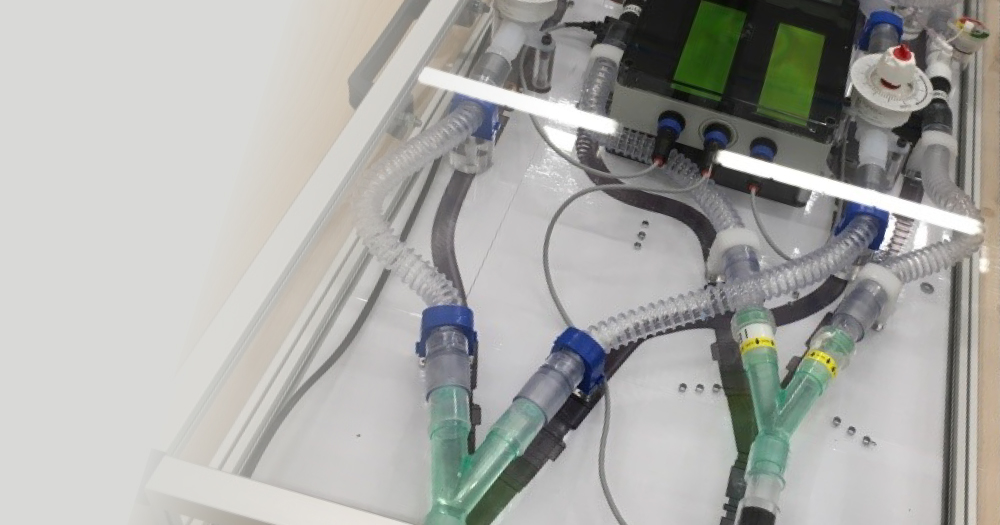
Website-Graphics_cdp-hero_ventilator-splitter-pr

New device could save lives around the world by allowing two patients to share one ventilator

Staff at Cambridge Design Partnership (CDP) have worked on an innovative project to “split” a medical ventilator, so that it can save the life of two patients at the same time. It could be used by healthcare providers around the world to double ventilator support in emergencies involving mass casualties. The innovative device is now set for regulatory approval thanks to the CDP team.

The device was initially created in a tight timeframe in March 2020, said Jon Cooke, who led CDP’s team working on the project. “At the time, there were fears that Royal Papworth Hospital in Cambridge could not cope with the high numbers of seriously ill Covid-19 patients they might face, so we worked round the clock to turn the idea into reality.”

The device allows one ventilator to serve two patients safely, even if they have differing needs for breathing support. It is now being tested and looks set to become a key piece of emergency equipment in pandemics and other crises such as war and mass shootings.

The project was initiated by the Institute for Manufacturing (IfM), University of Cambridge, in response to a call for help from Royal Papworth Hospital anaesthetists. Previous ventilator-splitting designs did not take into account the differing needs of the two patients in terms of lung capacity and breathing needs. “They were so rudimentary they ran the risk of doing serious damage to the patients’ lungs,” said Jon.

All parts in the device are easily sourced and can be swiftly changed and replaced. Initial details of how to build the device are now freely available on the Institute for Manufacturing website, for use by all medical professionals in emergencies. “If other communities across the world have to consider this emergency option in the event of a shortage of ventilators, they can copy and adapt the set-up for their own needs,” said Jon.

“The CDP team was asked to join the project at short notice over Easter 2020. “Although ventilator demand in the UK has now reduced, this system could provide emergency support in other countries which are still facing significant challenges with the pandemic. It’s also of great help for longer-term use in countries with ongoing ventilator shortages,” said Jon Cooke.

Two anaesthetists from the Royal Papworth Hospital, Professor Andrew Klein and Dr Chinmay Patvardhan, initiated the project. Professor Klein said: “We needed to have the ability to measure and control the air flow to each patient individually. We also needed to ensure that if there was a decline or improvement in breathing in one patient this would have no effect on air delivery to, or monitoring of, the other.”

“It was also essential that the set-up was easy to assemble and use. The device is currently being tested at Royal Papworth Hospital and first trials of the device using artificial lungs are very encouraging.”

Dr Patvardhan said: “This device could allow medical professionals to instantly double their ability to ventilate patients safely in a crisis situation. This would be invaluable for any future emergency on the scale we have seen with COVID-19.”

The system is specifically designed to be simple to use and maintain. The unit provides isolated respiratory lines to two patients and can be swiftly added to a ventilator. A meter shows a measurement of tidal volume to each patient. The device can also monitor the total pressure and airflow in the device. Easy-to-access valves enable fine tuning of air flow to match the requirements of each patient.

Jon Cooke said: “It was a hugely positive experience for us here at CDP to support the IfM team and work with Royal Papworth to deliver a functioning and safe design in such a short space of time.

“The combination of the deep regulatory knowledge and robust design techniques of the CDP team along with the great conceptual and experimental work conducted by the IfM made for a truly rapid development process. I’m extremely proud of what we achieved together in this remarkable cooperative project. “Of course, we hope that such emergency use of ventilators will be a rare occurrence in the future. But if such a situation arises, this system can be a lifesaver.”


Watch a report on the new device from BBC Science and Technology Correspondent Richard Westcott:


‘Invaluable’ ventilator device could treat two Covid-19 patients at once

BBC News coverage

Royal Papworth tests new ventilator for use on two patients simultaneously

ITV News coverage


For more information on the ventilator sharing device project, visit the Institute for Manufacturing (IfM) website here.

For further information and media enquiries, please contact: media@cambridge-design.com or call 01223 264428